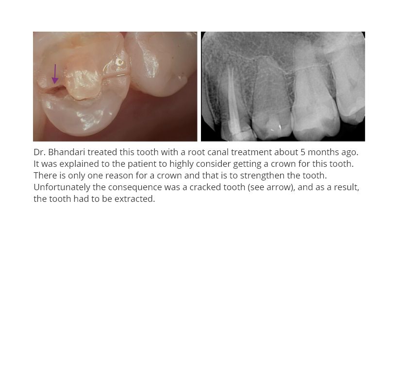
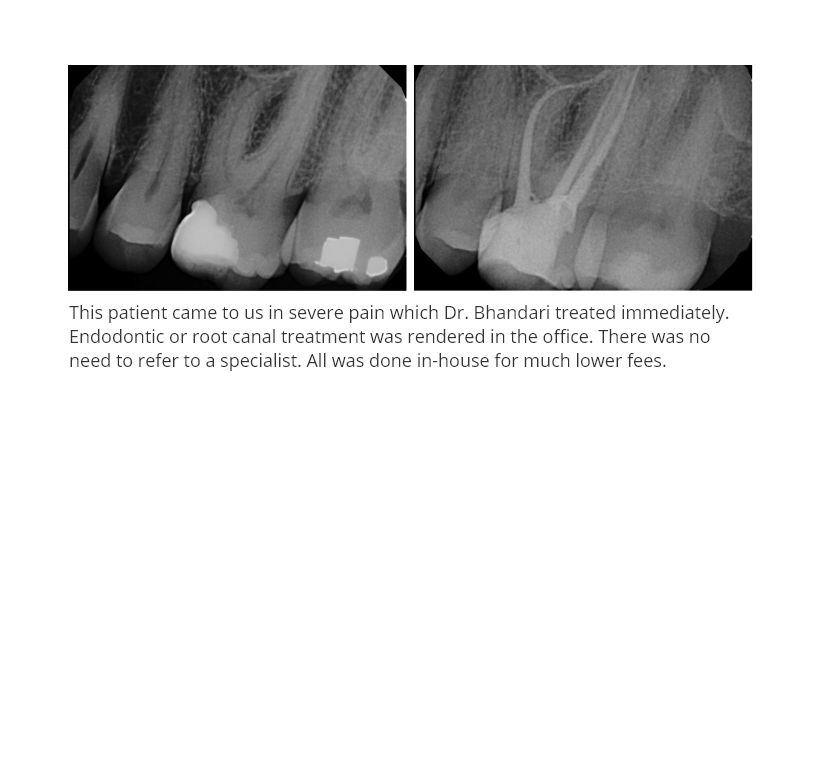
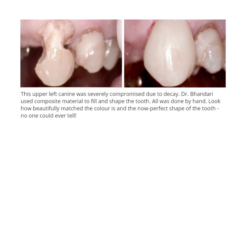
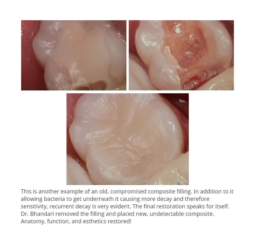
Dental radiographs can provide important information about your oral health. The
radiographs also become an essential part of your dental records. Some of things that they help diagnose include small area of decay between the teeth which are not otherwise visible to the naked exam, some types of tumors, cysts, bone loss due to periodontal disease or bone destruction due to carcinomas or tooth abscesses, position of un-erupted teeth in children, developmental abnormalities etc etc.
Digital radiography like the one we use in our offices uses a radiography machine and a small electronic sensor that is placed in your mouth to capture the image. The image is transmitted to a computer which than shows the image. The radiographic image can be viewed both on a computer monitor or a large TV screen for the patient to see.
Conventional X Rays usually take pictures on a larger area of the body while as Dental X Rays take pictures of a very small targeted area. The use of a cone to take a picture combined with a small target and the use of a lead apron with a thyroid collar makes it very safe and the information gained is invaluable.
According to the American and Canadian Dental Association, the radiation you receive from digital radiography is very small to minuscule. The millisievert (mSv) is the scale used to measure radiation. You could be exposed to .038 mSv for 2 Bitewing Digital Radiographs and .150 mSv from a Full Mouth Series (about 18 photos).
When you compare this to
CT Angiogram 16mSv
CT Chest 7mSv
CT GI Tract 5mSv
FULL MOUTH DENTAL DIGITAL RADIOGRAPH (18 pictures) 0.15mSv
Average natural radiation exposure in North America per year is 3.0mSv
One plane trip exposes you to .03mSv
The recommended maximum whole body radiation exposure per year is 50mSv (10mSv=1Rem)
As you can see the radiation exposure from digital dental radiographs is negligible. You are exposed to more radiation by just surviving than you are by going for a dental visit. But the benefits from dental digital radiography are huge.
Some of the Digital Radiography benefits include:
1)Higher Quality of Care-the equipment you use shapes the way your patients perceive your practice. Digital radiography reduces the exposure between 80-90%
2)No Chemicals for Developing-there is no more harmful chemicals to use and store. There is no more wait time for development of the picture taken and of courses no more odour.
3)Referring and Sharing of Information-it allows us to email images to specialists, and or benefit companies in no time. There is better discussion with a colleague when both are looking at the same image.
3)Image Enhancement-you can control the size, density, color code, label, place arrows etc which allow you to better diagnose and once again communicate with others on the case. You can also share this image with the patient for great patient involvement and treatment acceptance.
4)Image Quality-the image is of a much higher quality and less likely to miss diagnose.
5)Saving Space-no more saving x-rays in the patients charts/file, it is all electronically stored on a disc in the computer. Access can be from anywhere in the office and from home if you are working on a file.
6)Production and Time Management-if the office requires you to send images to your insurance company for a pre-determination or to a specialist you don’t need to make duplicates and mail them, both time and cost saving.
7)Lost X-Rays-no more lost images from the chart or from the mail, it is all electronic. All data is backed up daily, hence no loss or missing images.
8)Lower dose of Radiation.-patients are very happy about this and share this information with their friends, neighbours and colleagues. In turn results in greater referral.
9)Easier and Faster Usage-it is much faster and easier to utilize digital radiography than the conventional x rays.
The only disadvantage is the initial start up cost. In my opinion the advantages out way the disadvantages by far. With this new advancement in technology, both the dentist andpatient can be involved in the treatment planning as a result of the patient observing the same thing the dentist sees. This leads to better care, patient acceptance, less radiation exposure and environmental damage as a result of no solutions and or chemicals involved. It’s a win- win situation.
BY Dr V Bhandari Professional Dentistry
905-902-9000
